


Diagnostic Ultrasound & Ultrasound-Guided Injections
Real-time evaluation and image-guided procedures for selected joint, tendon, muscle, soft-tissue, nerve, and spine-related pain conditions.
Diagnostic ultrasound can help evaluate selected muscles, tendons, joints, bursae, superficial nerves, blood vessels, soft tissues, and certain spine-related structures in real time. When an injection is appropriate, ultrasound guidance allows the physician to visualize the target and nearby anatomy during the procedure.
At Network Spine, ultrasound is used as part of a broader physician-led evaluation that may include osteopathic assessment, palpation, imaging review, fluoroscopic guidance, and functional examination. The goal is not simply to see anatomy, but to determine whether a finding is clinically relevant.
What Diagnostic Ultrasound Can Evaluate
Diagnostic ultrasound uses sound waves to evaluate selected musculoskeletal and soft-tissue structures in real time. In medical settings, this may also be called diagnostic musculoskeletal ultrasound or MSK ultrasound.
Depending on the clinical situation, ultrasound may help evaluate:
Tendons
Muscles
Ligaments
Nerves
Bursae
Blood vessels
Joints and joint fluid
Soft-tissue swelling or fluid collections
Spine and spine-adjacent structures
Areas of focal tenderness or symptom reproduction
Dynamic assessment during movement, contraction, stress, or direct pressure
One advantage of ultrasound is that it allows dynamic assessment. Some structures can be evaluated during motion, contraction, pressure, or direct palpation with the ultrasound transducer. This can help determine whether a visible finding is clinically relevant or simply an incidental structural abnormality.
Ultrasound does not replace MRI, X-ray, electrodiagnostic testing, fluoroscopy, or other diagnostic studies when those are clinically necessary.
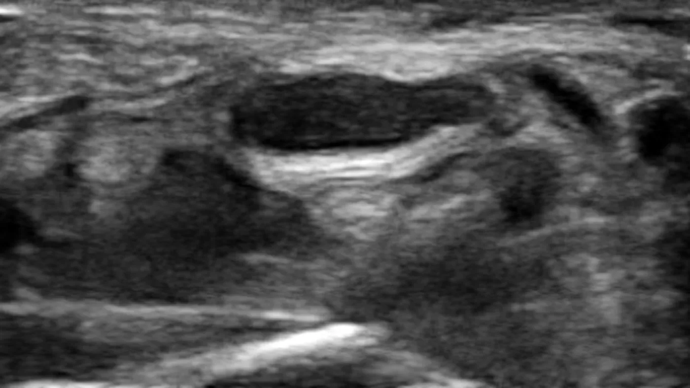
Diagnostic ultrasound can help evaluate selected superficial nerves and soft-tissue structures in real time.
Sonopalpation: Combining Ultrasound and Examination
A valuable technique used at Network Spine is sonopalpation. During sonopalpation, live ultrasound imaging is combined with direct pressure from the ultrasound transducer. This allows the physician to visualize a structure while also determining whether pressure over that structure reproduces the patient’s familiar pain.
This distinction is clinically important. Imaging can show structural changes that may or may not be responsible for symptoms. Palpation can help identify whether a structure is actively painful. When the two are combined, concordant pain reproduction may improve diagnostic confidence and procedural planning.
Sonopalpation can be especially useful when evaluating soft-tissue pain, trigger points, tendon-region pain, bursa-related pain, peripheral nerve irritation, and selected spine-adjacent pain patterns.
Ultrasound-Guided Injections
Ultrasound guidance may be used for selected injections involving soft tissue, tendon regions, bursae, joints, muscles, superficial nerves, nearby blood vessels, and certain spinal or spine-adjacent structures.
Depending on the diagnosis and clinical plan, ultrasound guidance may be considered for:
Trigger point injections
Joint injections
Bursa injections
Tendon injections
Peripheral nerve blocks
Selected soft-tissue injections
Selected spine injections
Orthobiologic procedures when appropriate
Ultrasound-guided injections are often paired with a broader treatment plan that may include osteopathic treatment, rehabilitation guidance, activity modification, medication when appropriate, or fluoroscopic procedures when a deeper spine target is suspected.
Related: Trigger Point Injections, Osteopathic Approach, Advanced Injection Options, Nerve Blocks
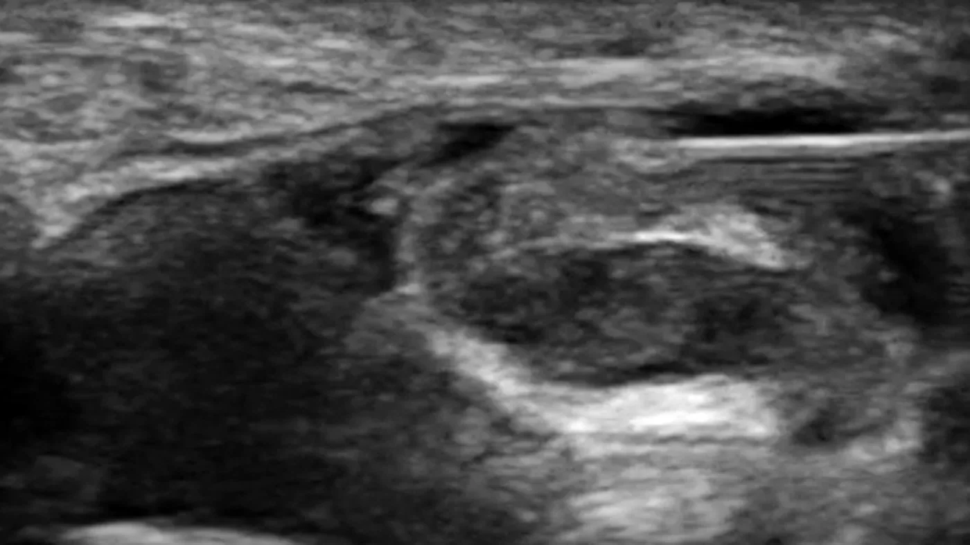
Ultrasound guidance provides real-time visualization of the target, needle path, and surrounding anatomy during selected injections.
Guidance Is More Than Imaging
Injection guidance is not limited to a single technique. Depending on the clinical situation, ultrasound, fluoroscopy, anatomic landmarks, and palpation may each play a role. In selected cases, these methods may also be combined.
Ultrasound is particularly useful when the target involves soft tissue, tendon, bursa, joint, muscle, superficial nerve, nearby blood vessels, or selected superficial spinal structures. It allows real-time visualization and can help identify structures that may not be well seen with fluoroscopy alone.
Fluoroscopy provides excellent visualization of bony landmarks and is often preferred for many deeper spine-related procedures, where contrast may be used to confirm spread patterns. In some cases, ultrasound and fluoroscopy may be complementary. For example, ultrasound may help identify superficial vascular structures before a procedure, while fluoroscopy may help confirm deeper bony or contrast-based landmarks.
Palpation remains clinically important, especially for trigger point injections and soft-tissue pain. It helps determine whether a structure is reproducing the patient’s symptoms rather than simply appearing abnormal on imaging.
The goal is not simply to place a needle near an anatomic structure. The goal is to determine whether that structure is clinically relevant.
Related: Epidural Steroid Injections, Facet Joint and Medial Branch Blocks, Sacroiliac Joint Injections, Radiofrequency Ablation
Ultrasound, Fluoroscopy, Landmarks, and Palpation
Different guidance methods serve different purposes.
Ultrasound may be useful for soft tissue, tendon, bursa, joint, muscle, superficial nerve, nearby blood vessels, and selected superficial spine-adjacent targets.
Fluoroscopy is often preferred for many deeper spine-related procedures, including epidural steroid injections, facet-related procedures, and some sacroiliac joint injections.
Anatomic landmarks may help guide certain procedures when the target is clinically straightforward and image guidance is not necessary.
Palpation may help identify focal tenderness, tissue texture change, trigger points, and symptom reproduction during examination.
These methods are not mutually exclusive. The appropriate guidance method depends on anatomy, safety considerations, the suspected pain generator, and the purpose of the injection.
Conditions and Body Regions Commonly Evaluated
Diagnostic ultrasound may help evaluate selected tendon, bursa, joint, muscle, and soft-tissue sources of shoulder pain. It may also be used to guide selected shoulder-region injections when clinically appropriate.
Ultrasound may be useful for selected anterior, lateral, and soft-tissue hip-region pain patterns. Some hip-region injections may be performed with ultrasound guidance, depending on the suspected pain source and anatomy.
Back, Neck, and Spine-Adjacent Pain
Many spine-related procedures are best performed with fluoroscopic guidance. However, ultrasound may be useful for selected superficial spinal or spine-adjacent structures, soft-tissue pain generators, trigger points, and peripheral nerve-related pain patterns. In some cases, ultrasound may complement fluoroscopy rather than replace it.
Certain superficial peripheral nerves can be evaluated or targeted with ultrasound depending on the clinical question. Ultrasound may help visualize nearby soft tissues, vascular structures, and selected nerve-region anatomy in real time.
Sports and Activity-Related Pain
Ultrasound may be useful for selected tendon, muscle, bursa, joint, and soft-tissue problems related to training, lifting, running, racquet sports, martial arts, or work-related activity. Its role is determined by the examination and the suspected pain generator.

Selective, Clinically Directed Use
Ultrasound findings are interpreted in the context of symptoms, examination findings, prior imaging, palpation, and functional goals. Not every structural finding is clinically relevant, and not every painful area requires an injection.
At Network Spine, ultrasound is used when it can add meaningful information to the evaluation or improve procedural accuracy. Treatment planning may include osteopathic treatment, rehabilitation guidance, medication when appropriate, image-guided procedures, fluoroscopic procedures, or referral when another type of care is more appropriate.
What a Visit Typically Involves
A visit generally begins with a focused history and physical examination. Prior imaging, prior treatments, activity demands, work demands, and functional limitations are reviewed when relevant.
When ultrasound is appropriate, it may be used to evaluate selected structures in the symptomatic region. In some cases, sonopalpation may be used to determine whether pressure over a visible structure reproduces the patient’s familiar pain.
If an injection is being considered, the physician will discuss the suspected pain generator, the rationale for the procedure, alternatives, expected limitations, and relevant risks. Some patients are better managed without an injection. Others may benefit from a targeted procedure when the clinical picture supports it.
Why This Approach Is Different
Many pain problems are not explained by imaging alone. A tendon, joint, disc, bursa, nerve, or muscle may look abnormal without being the primary source of symptoms. Conversely, a clinically important pain generator may not be obvious without careful examination, palpation, dynamic assessment, and procedural judgment.
Network Spine combines osteopathic examination, diagnostic ultrasound, sonopalpation, fluoroscopic procedure experience, and interventional pain training within a single physician-led evaluation. This allows symptoms to be assessed across multiple possible pain sources rather than through a single imaging method or protocol.
Related: Physician Credentials
Frequently Asked Questions
Is diagnostic ultrasound the same as MRI?
No. Ultrasound and MRI provide different types of information. Ultrasound can evaluate selected structures in real time and may allow dynamic assessment. MRI may be preferred for deeper structures, complex anatomy, surgical planning, or conditions not well evaluated by ultrasound.
Does ultrasound guidance make injections more accurate?
For many soft-tissue, tendon, bursa, joint, muscle, superficial nerve, and selected superficial spine-adjacent targets, ultrasound guidance can improve anatomic accuracy by allowing the physician to see the target and nearby structures in real time. Some procedures are difficult, less precise, or not appropriate to perform without image guidance.
That said, ultrasound is not the best tool for every injection. Fluoroscopy is often preferred for deeper spine procedures, including many epidural, facet-related, and sacroiliac joint injections. The best guidance method depends on the anatomy, safety considerations, and clinical objective.
What is sonopalpation?
Sonopalpation combines live ultrasound imaging with direct palpation using the ultrasound transducer. It may help determine whether pressure over a visible structure reproduces the patient’s familiar pain, which can add diagnostic and procedural precision in selected cases.
Can ultrasound show nerves?
Certain superficial peripheral nerves can be evaluated with ultrasound depending on the region and clinical question. Some nerve-related symptoms may also require MRI, electrodiagnostic testing, or other evaluation.
Why use palpation if imaging is available?
Palpation can help identify whether a structure is clinically active and reproduces the patient’s symptoms. This is especially important for trigger points, soft-tissue pain, and some tendon or nerve-region complaints.
Do I need an injection if ultrasound shows an abnormality?
Not necessarily. Many imaging findings can be incidental. Treatment depends on whether the finding fits the history, physical examination, functional limitation, and overall clinical picture.
Is this a general radiology ultrasound service?
No. This page refers to diagnostic ultrasound used as part of a physician-led musculoskeletal, sports medicine, spine, and pain evaluation. It is not a general ultrasound service for abdominal, vascular, cardiac, pregnancy-related, or unrelated medical imaging.
Request an Evaluation
Diagnostic ultrasound and ultrasound-guided injections may be considered when they add meaningful information to the clinical evaluation or improve procedural accuracy. The appropriate approach depends on the suspected pain source, anatomy, prior treatment history, and examination findings.